
Profile
Laura Devlin
My CV
-
Education:
Hummersknott School, Darlington
Queen Elizabeth Sixth Form, Darlington
Durham University
Newcastle University
-
Qualifications:
GCSE: Triple Science (Biology, Chemistry, Physics), Maths, English, English literature, Geography, Performing Arts.
A-level: Biology, Chemistry, Maths, Geography, Critical Thinking.
BSc Biological Sciences
MRes Medical Genetics
PhD Medical Genetics…ongoing..
-
Work History:
Part time jobs:
1) Waitress
2) Retail assistant
3) Dance teacher -
Current Job:
PhD Medical Genetics
-
About Me:
Hi 👋 I’m Laura! I am a PhD student 👩🏻🔬, DNA fan 🧬 and dance addict 💃! I’m also a lover of chocolate 🍫!!
-
Read more
Hi everyone 👋, I’m Laura! I am currently in the 3rd year of my PhD at Newcastle University, to become a Dr of Medical Genetics 🧬! When I am not in the lab 🔬 I like to spend my time doing ballet, tap and jazz dancing 💃. I also like watching my favourite tv show…..Strictly Come Dancing! (I wonder if one day, they will let scientists dance on Strictly, instead of celebrities?) I also enjoy cycling 🚴♀️ and reading 📖. I love sweet treats, my favourites are red velvet cake 🍰 and creme eggs (I still have a few secretly hidden around the house…but don’t tell my brother!)
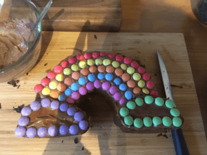
This is one of my more recent cake creations! (Although I eat more cake then I actually bake!)
-
Read more
I work on rare diseases known as ciliopathies, pronounced ‘silly-o-path-ees’ 😃. These are genetic diseases caused by ‘errors’ in DNA, which is the genetic material in our body that makes us all unique!
Unfortunately, people with these diseases may get very sick, and might have problems with their kidneys, eyes 👀 and brain 🧠.
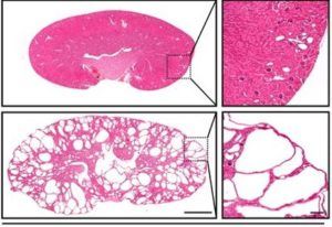
These are mouse kidneys. The top image demonstrates a very mild cystic kidney disease, with small cysts (holes) in the kidney. The bottom left image shows a severe form of the cystic kidney disease, look how big the cysts (holes) are! Unfortunately this bottom kidney does not function well, and is similar to the kidneys seen in some human ciliopathies. (Ramsbottom et al, 2020).
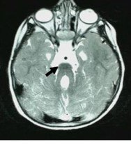
This is a magnetic resonance image (MRI) of a human brain. The black arrow is pointing to the centre of the brain, which has a grey shape that looks a bit like a molar tooth. We call this the ‘molar tooth sign’, and it is often found in the brains of patients with a certain type of ciliopathy. This can cause a range of problems including problems with speech, balance and learning. (Ramsbottom et al, 2018)
People who have these diseases have a faulty primary ‘cilia’, which is the tv aerial of our body’s cells, that pick up signals, telling each of our cells what to do. 📺
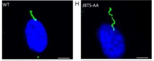
These are kidney cells (renal epithelial). In blue is the centre of the cell, it’s nucleus. In green we can see the amazing cilia, sticking out of the cell, ready to detect signals. On the left is a normal cell, on the right is a faulty cell. What differences can you see in the cilia? (Ramsbottom et al, 2018)
My job is to understand..
1) Why do problems with the primary cilia cause people to get sick, and can we treat them?
2) What DNA ‘errors’ cause these diseases?
To investigate this I use mice, cells and genetic data. The mice I use have a genetic ‘mutation’, similar to the error seen in the humans with some ciliopathy diseases. I use them to understand what goes wrong when you have this genetic error, and if there are ways of treating the disease.
Our lab also has a special way of extracting kidney cells from peoples wee, and then we can look at cells under a microscope to see if there is anything wrong, and test whether some medicines can make them better.
I also search the DNA of patients, to see whether the ‘error’ causing the disease can be found.
-
My Typical Day:
I snooze my alarm many times (because I like my sleep 😴), before getting out of bed, grabbing breakfast and hopping on a train 🚂 to work. I grab a quick tea ☕️ when I get into work, say hi 👋 to all my colleagues and check my plans for the day, then off to the lab 👩🏻🔬 🧪 🧫 🔬 I go!
-
Read more
Once I get to work, for about 9am, I get myself a tea ☕️ sit down at my computer 💻 , and read all my emails 📧 , answering any needing immediate attention. I check my calendar 🗓 where I have planned all my experiments for the week, and then I get down to the lab for about 9:30 👩🏻🔬.
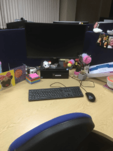
This is my desk in the office. I like to make it as fun as possible, I have colourful sticky notes, pictures and most importantly, lots of fluffy pens!
I can spend the morning in the lab, doing anything from genotyping DNA, staining tissues/cell samples with fluorescent markers or histological dyes, as well as going to see my mice! My mice are looked after by a specialised team of experts, and vets, who make sure they are all in tip top condition, get all the food and water they need, and have lots of forms of mental stimulation. At the end of the experiment, I have to do a mouse dissection, so I can look at their kidneys, eyes, brain and other tissues, to see what is wrong.
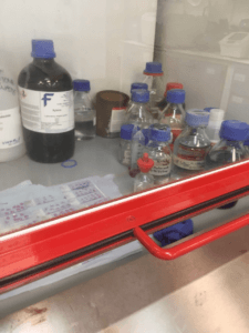
Staining equipment used to stain thin slices of mouse kidney a pink colour, so I can see the cells and structure. This is done inside a fume hood, so I do not breathe in the toxic fumes.
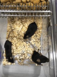
These are some of my mice. Mine all have black fur. Have you noticed the little hole in the ear of the mouse at the front….what do you think this is for?
At about 12:30 I have lunch 🥙 with my friends, colleagues and fellow students (normally a sandwich….but I have a Greggs if I’m treating myself!) and we chat about our day, tv, the news….anything and everything!
Twice a week we have a seminar at 13:00 for about 45 minutes, where students and staff can talk about their current research. It’s a great chance to learn things outside your expertise!
Once a week we also have a meeting with my whole lab group, to discuss our plans for the week and help each other with any problems.
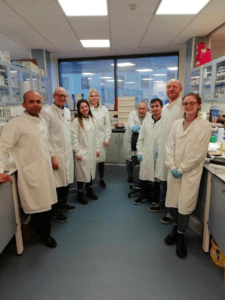
This is a picture of my lab group. We work together on research projects, guided by my supervisors John, Colin and Simon.
I then often go back to the lab, to finish off the day’s experiment, go on the microscope 🔬 to take pictures of cells and tissues, or make up reagents for tomorrow’s experiment.
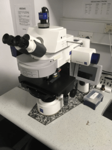
This is one of our microscopes, that we use to take images of tissues and cells with fluorescent markers. When we use this microscope we have to sit in the dark, so we don’t spoil the images!
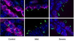
This is an example of some of the images we can see on the immunofluorescence microscope. This is a small section of mouse kidney tissue. In green we can see the cilia, in blue we can see the individual cells. “Control” is the normal kidney. As we move right we can see that the kidney cilia get longer and curly, but it also loses it’s pink stain (for a protein called Barttin). (Ramsbottom et al, 2020)
At around 3:30pm, I go back to the office and grab a snack, and then I look at some of the microscope images I have taken, analyse any experimental data or look at some patients DNA sequences. I often spend time writing ‘papers’ which are reports of my research, and preparing talks to give to people at difference conferences.
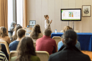
This is me, presenting my research to a group of students. I am lucky that as a scientist I get to travel to different places and talk about my work, as well as meet lots of other scientists!
Importantly, my day is never the same, which makes my job so great!
-
What I'd do with the prize money:
I’d like create a science podcast for early career researchers like me, to talk about their specialist subject, getting more people interested in science!
-
My Interview
-
How would you describe yourself in 3 words?
Smiley, Curious, Unique
What did you want to be after you left school?
I had no idea!
Were you ever in trouble at school?
Not really
Who is your favourite singer or band?
Beyoncé
What's your favourite food?
Chocolate!!!!
If you had 3 wishes for yourself what would they be? - be honest!
1) be healthy 2) be happy 3) ......get a dog!!!
-